
Overview about Centrioles
Presence is found in animals, microbes, and lower plants. These are present at a right angle to each other.
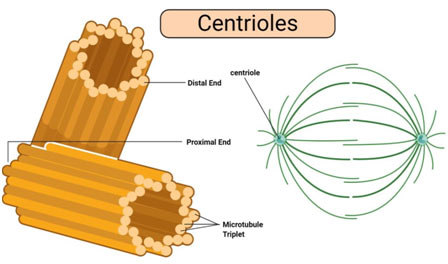
- The cross-section shows an array of 9 microtubules, each composed of 3 tubules called triplet of tubules.
- These microtubules radiate from the center.
- Before cell division, duplication of centrioles takes place and each one moves to opposite poles developing spindle fibers.
- Centrioles control chromosome movement, and also organize the cytoskeleton.
- They decide the location of furrowing during cell division. Centrioles also help in the formation of cilia.
- 1) Centrioles
- 2) Structure of Centrioles
- 3) Distal Part
- 4) Central Core
- 5) Centriole Duplication
- 6) Functions of Centrioles
- 7) Centriole in Plants
- 8) Multiple Choice Questions (MCQs) with Answers
- 9) Frequently Asked Questions (FAQs)
- 10) Tutorial Summary: Centrioles – Structure, Functions & Centriole in Plants
- 11) You may also like to learn:
Centrioles
Eukaryotic cells include two round, rod-shaped, microtubular structures, called centrioles, near the nucleus.
They do not have a restricting membrane and DNA or RNA and take place in a lot of algal cells (a notable exception being red algae), moss cells, some fern cells, and a lot of animal cells. They are missing in prokaryotes, red algae, yeast, cone-bearing and flowering plants (conifers and angiosperms), and some non-flagellated or non-ciliated protozoans (such as amoebae).
Centrioles form a spindle of microtubules, the mitotic device during mitosis or meiosis and in some cases get set up just underneath the plasma membrane to form and bear flagella or cilia in flagellated or ciliated cells. When a centriole bears a flagellum or cilium, it is called the basal body.
Structure of Centrioles
With a diameter of about 250nm and a length ranging from 150-500nm in vertebrates, centrioles are some of the largest protein-based structures. The nine triplets microtubules are some of the most recognizable functions of this organelle.
In some organisms (e.g. in Drosophila and nematodes) the microtubules are simpler and may occur as either doublet microtubules (in flies) or single microtubules as holds true with Caenorhabditis elegans.
In human beings, however, among other higher animals, they exist as intricate triplets that make up the scaffold of the microtubules arranged in a circle (at an angle) around the central core. When seen from one end, the triplet microtubules appear to have an anticlockwise twist plan.

Essentially, the centriole is composed of three main parts. These consist of:
Distal Part
The distal part of the centrioles is identified by the microtubules (triple or double). This part is also divided into the distal and sub-distal parts/appendages. While eukaryotic cells consist of an overall of nine distal appendages, sub-distal appendages differ in number depending on cell type and functions.
Structure-wise, the distal appendages look like turbine blades that are symmetrically set up at the distal end of the centriole. Here, each of the appendages is connected to one of the triplets at 50 degrees angle to the centriole surface.
Unlike the distal appendage, sub-distal appendages are attached to two or three triplets and form an aright angle with the centriole surface. Sub-distal appendages have likewise been revealed to alter shape/morphology and even disappear in some cases. Apart from differences in shape/morphology and plan, distal and sub-distal appendages also have different functions.
For instance, the distal appendages serve to connect the centriole during cilium development in some cells, whereas sub-distal appendages function as centers of nucleation for microtubules.
Central Core
The central core is the part of a centriole on which microtubule triplets are connected. In such organisms as C. reinhardtii, this structure is about 250nm in length and has a Y-shaped linker in addition to a barrel-like structure situated in its inner core. As part of the centriole, the central core serves to support the scaffold.
The adjacent triplet fibrils are linked by– proteinaceous linkers. The center of the centriole has a rod-shaped proteinaceous mass called the hub. The hub has a diameter of 2.5 nm. From the hub, develops 9 proteinaceous hairs towards the peripheral triplet fibrils.
They are called spokes. Each spoke has actually a thickening called X before uniting with A sub-fiber. Another thickening called Y exists close by. It is connected both to X thickening in addition to C– A linkers by connectives. Due to the presence of radial spokes and peripheral fibrils, the centriole gives a cartwheel look in T.S.
Centriole Duplication
Like chromosomes, centrioles also duplicate during cellular division. Although it was believed that a new daughter centriole was the product of the pre-existing centriole (functioning as the template for the new centriole), studies have shown following over-expression of centriolar proteins, new centrioles can be formed. For this factor, new centrioles do not always originate from pre-existing centrioles.
However, in a variety of scientific research studies where pre-existing centrioles were entirely removed, duplication was also impacted. Regardless, just a single new centriole is produced with every cell cycle. New/ daughter centrioles are generally assembled during the S stage of the cell cycle.

Functions of Centrioles
- Cells form a complicated endoskeleton of microtubules which permits substances to be transferred to any place in a cell. Products are tagged with unique glycoproteins (sugar and protein) which function as signals to particular motor proteins. These proteins connect to the product, or vesicle that the product is saved in, and also attach to a microtubule. Microtubules are organized at the centriole, of which each centrosome has 2. The centrioles anchor the microtubules that extend from it and consist of the elements needed to produce more tubules
- Throughout mitosis, centrosomes are replicated by duplicating each centriole. The 4 centrioles then divide into 2 centrosomes, each with one centriole at a right angle to the second centriole. Microtubules extend between the centrosomes which push the sets of centrioles apart. The centrioles will be pushed apart, to opposite ends of the cell. Once established, each centriole will then extend microtubules into the cytoplasm that seek out chromosomes. The microtubules connect to the chromosomes at their centromeres, which are parts of the DNA specifically created to enable the attachment of unique proteins and microtubules. The microtubules are then disassembled from the centriole, which draws the microtubule back towards the centriole, as motor proteins pull the chromosomes apart.
- The absence of centrioles causes divisional errors and delays in the mitotic process.
- A single centriole forms the anchor point, or basal body, for each individual cilium or flagellum.

- Basal bodies direct the development of cilia and flagella too.
Centriole in Plants
Higher plants do not have centrioles. Spindle fibers that help with the separation of chromosomes are for that reason produced by an organelle referred to as centrosome. While the organelle is lacking in greater plants, it can be discovered in some lower plants. For example, in such lower plants like mosses, ferns, and cycads, centrioles have actually been shown to form during spermatogenesis (a form of cell division).
Multiple Choice Questions (MCQs) with Answers
1. What is the main function of centrioles?
- a) Synthesizing DNA
- b) Controlling chromosome movement
- c) Producing ATP
- d) Storing lipids
Answer: b) Controlling chromosome movement
2. In which cells are centrioles typically found?
- a) All eukaryotic cells
- b) Prokaryotic cells
- c) Red algae cells
- d) Plant cells
Answer: a) All eukaryotic cells
3. What is the structure responsible for supporting the scaffold in the centriole called?
- a) Central Core
- b) Distal Appendage
- c) Sub-Distal Appendage
- d) Hub
Answer: a) Central Core
4. How are the microtubules arranged in the centriole?
- a) Singlet microtubules
- b) Doublet microtubules
- c) Triplets arranged in a circle
- d) Quadruplet microtubules
Answer: c) Triplets arranged in a circle
5. During which stage of the cell cycle does centriole duplication occur?
- a) G1 phase
- b) S phase
- c) G2 phase
- d) M phase
Answer: b) S phase
6. What is the role of distal appendages in the centriole?
- a) Anchoring microtubules
- b) Initiating cell division
- c) Connecting the centriole during cilium development
- d) Nucleation for microtubules
Answer: c) Connecting the centriole during cilium development
7. Which part of the centriole is characterized by turbine blade-like structures?
- a) Distal Part
- b) Central Core
- c) Centriole Duplication
- d) Basal Body
Answer: a) Distal Part
8. What is the diameter range of centrioles in vertebrates?
- a) 50-100nm
- b) 150-500nm
- c) 700-1000nm
- d) 1000-1500nm
Answer: b) 150-500nm
9. In plant cells, what organelle substitutes for centrioles in organizing spindle fibers?
- a) Chloroplast
- b) Centrosome
- c) Mitochondrion
- d) Nucleolus
Answer: b) Centrosome
10. What happens to centrioles during mitosis?
- a) They disintegrate
- b) They duplicate
- c) They remain inactive
- d) They migrate to the nucleus
Answer: b) They duplicate
11. What is the function of centrioles during cilium and flagellum development?
- a) Synthesizing microtubules
- b) Anchoring chromosomes
- c) Serving as basal bodies
- d) Initiating cell division
Answer: c) Serving as basal bodies
12. Which plant cells lack centrioles?
- a) Moss cells
- b) Fern cells
- c) Flowering plant cells
- d) Algal cells
Answer: c) Flowering plant cells
13. How are distal and sub-distal appendages different?
- a) They have different shapes
- b) They have different functions
- c) They are not connected to triplets
- d) Both a and b
Answer: d) Both a and b
14. What does the central core of the centriole support?
- a) Triplets microtubules
- b) Distal appendages
- c) Sub-distal appendages
- d) Basal bodies
Answer: a) Triplets microtubules
15. How does centriole duplication occur in the cell cycle?
- a) During G1 phase
- b) During G2 phase
- c) During M phase
- d) During S phase
Answer: d) During S phase
16. What happens if centrioles are absent during cell division?
- a) Normal division
- b) Divisional errors and delays
- c) Faster division
- d) Incomplete division
Answer: b) Divisional errors and delays
17. What is the structure at the end of each centriole that appears like an anticlockwise twist?
- a) Distal appendage
- b) Hub
- c) Sub-distal appendage
- d) Basal body
Answer: a) Distal appendage
Frequently Asked Questions (FAQs)
1. What is the main function of centrioles?
- Centrioles control chromosome movement, organize the cytoskeleton, decide the location of furrowing during cell division, and contribute to the formation of cilia.
2. Where are centrioles typically found in cells?
- Centrioles are found in eukaryotic cells, specifically in animals, microbes, and lower plants. Notable exceptions include red algae, yeast, cone-bearing and flowering plants, and some non-flagellated or non-ciliated protozoans.
3. What is the structure of centrioles?
- Centrioles are round, rod-shaped, microtubular structures with a diameter of about 250nm and a length ranging from 150-500nm in vertebrates. They consist of nine triplets of microtubules arranged in a circle around the central core.
4. What is the significance of the distal part of centrioles?
- The distal part is characterized by turbine blade-like structures called distal appendages. They connect the centriole during cilium development and play a role in organizing nucleation for microtubules.
5. How does centriole duplication occur?
- Centrioles duplicate during cellular division, and studies have shown that new centrioles can be formed independently of pre-existing ones. A single new centriole is generally assembled during the S stage of the cell cycle.
6. What functions do centrioles perform during mitosis?
- Centrioles are replicated during mitosis, and microtubules extend between the centrosomes, pushing the sets of centrioles apart. They anchor microtubules that seek out chromosomes, contributing to proper chromosome separation.
7. What happens if centrioles are absent during cell division?
- The absence of centrioles can lead to divisional errors and delays in the mitotic process.
8. Do plant cells have centrioles?
- Higher plants do not have centrioles. The spindle fibers that aid in the separation of chromosomes in plant cells are produced by an organelle called the centrosome, which is absent in higher plants.
9. What is the role of centrioles in cilium and flagellum development?
- Centrioles serve as basal bodies, directing the development of cilia and flagella.
10. How do centrioles contribute to the endoskeleton of microtubules in cells?
- Centrioles anchor microtubules and contain the elements needed to produce more tubules, allowing substances to be transported within the cell.
Tutorial Summary: Centrioles – Structure, Functions & Centriole in Plants
This tutorial provides a comprehensive understanding of centrioles, covering their structure, functions, and the absence of centrioles in plant cells. Here’s a brief summary of key points:
1. Overview:
- Centrioles are present in animals, microbes, and lower plants, arranged at right angles to each other. They consist of nine microtubules radiating from the center, playing a crucial role in cell division.
2. Centrioles:
- Found in eukaryotic cells, centrioles are round, rod-shaped structures near the nucleus. They form a spindle of microtubules during mitosis or meiosis and can act as basal bodies for flagella or cilia.
3. Structure of Centrioles:
- Centrioles have a diameter of about 250nm and a length ranging from 150-500nm. They consist of nine triplets of microtubules, forming a scaffold arranged in a circle around the central core.
4. Distal Part:
- The distal part includes distal and sub-distal appendages. Distal appendages connect the centriole during cilium development, while sub-distal appendages serve as centers of nucleation for microtubules.
5. Central Core:
- The central core is the part of the centriole where microtubule triplets are connected. It has a Y-shaped linker, barrel-like structure, and radial spokes, giving the centriole a cartwheel appearance.
6. Centriole Duplication:
- Centrioles duplicate during cellular division, with studies showing that new centrioles can form independently of pre-existing ones. A single new centriole is produced in each cell cycle during the S stage.
7. Functions of Centrioles:
- Centrioles anchor microtubules, facilitating substance transport within cells. During mitosis, they play a crucial role in chromosome separation. The absence of centrioles leads to divisional errors.
8. Centriole in Plants:
- Higher plants lack centrioles; instead, spindle fibers aiding chromosome separation are produced by the centrosome. Some lower plants, such as mosses and ferns, have been shown to form centrioles during spermatogenesis.
This tutorial offers a detailed exploration of centrioles, shedding light on their intricate structure, diverse functions, and the unique characteristics observed in different organisms.