The Human Eye
Our eyes are located in small portions of the skull known as the orbits or eye sockets. Eyelids wipe eyes and prevent dehydration.
They spread tears in the eyes, which include substances for combating bacterial infections. Eyelashes prevent large particles from getting into the eye.
Structure of Eye
The structure of the eye can be divided into three main layers.
External layer
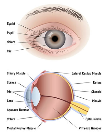
The outer layer of eyeball includes sclera and cornea. Sclera gives the eye the majority of its white colour. It consists of thick connective tissue and protects the inner components of the eye and preserves its shape. In the front, sclera forms the transparent cornea. Cornea admits light to the interior of the eye and flexes light rays so that they can be brought to a focus.
Middle layer
The middle layer is called the choroid. It consists of blood vessels and offers the inner eye a dark colour. The dark colour avoids disruptive reflections within the eye. Behind the cornea, choroid bends to form a muscular ring, called iris. There is a round hole, called pupil, in the centre of iris.
After striking the cornea, light travel through the pupil. The size of the pupil is changed by the muscles of iris. The pupil constricts in bright light when the circular muscles of the iris contract. Similarly, the pupil dilates in dim light when the radial muscles of the iris contract.
Behind iris, there is a convex lens, which focuses light on the retina. Lens is attached to ciliary muscles of the eye via a ring of the suspensory ligament. To clearly see an object far, ciliary muscles are relaxed and the lens becomes less convex. When ciliary muscles contract, the lens ends up being more convex and rounder.
Inner layer
The inner layer is sensory and is called the retina. It consists of the photosensitive cells called rods and cones and associated nerve cells.
Rods are sensitive to dim light while cones are sensitive to bright light and so identify different colours. Retina has two points i.e., fovea and optic disc. The fovea is a dip in the retina, directly opposite to lens and is largely packed with cone cells. It is largely responsible for colour vision and sharpness. The optic disc is a point on the retina where the optic nerve enters the retina. There are no rods and cones at this point, that is why it is likewise described as the blind spot.
The iris divides the cavity of the eye into two chambers. The anterior chamber is in front of iris i.e., between cornea and iris; whereas the posterior chamber is between iris and retina. The anterior chamber contains a clear fluid known as aqueous humour while the posterior chamber includes a jelly-like fluid known as vitreous humour. It assists keep the shape of the eye and suspends the delicate lens.
Light from objects enter into the eye and is refracted when it travels through the cornea, aqueous humour, lens and vitreous humour. Lens likewise focuses light on the retina. As a result, the image falls on the retina. Rods and cones produce nerve impulses in the optic nerve. These impulses have reached the brain, that makes the experience of vision.
Rods consist of a pigment called rhodopsin. When light falls on rhodopsin, it breaks for producing a nerve impulse. In the lack of light, the breakdown products are once again converted into rhodopsin. The body synthesizes rhodopsin from vitamin Aand that is why the shortage of vitamin A causes poor night vision. This issue is called night blindness.
Cones likewise include a pigment, referred to as iodopsin. There are three main types of cones and each type has a particular iodopsin. Each type of cones acknowledges among the three primary colours i.e., blue, green and red. If any kind of cones is not working well, it becomes challenging to acknowledge that colour. Such a person is likewise unable to differentiate different colours. This disease is called colour blindness and it is a genetic issue.
Disorders of the Eye
The working of the eye is affected by the changes in the shape of the eyeball.
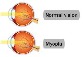
Myopia (Short sight)
The elongation of eyeball leads to myopia. Such persons are not able to see far-off things clearly. The image of a remote object is formed in front of the retina. This problem can be rectified by using a concave lens.
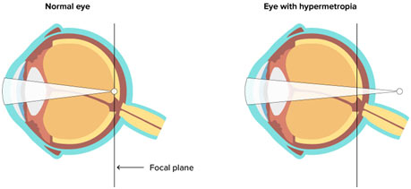
Hypermetropia (Long sight)
It takes place when eyeball reduces. Such individuals are not able to see near objects clearly. The image is formed behind the retina. The convex lens is used to rectify this issue.
MCQs – Human Eye – Structure and Disorders of the Eye
- What is the function of the eyelids in the human eye?
- a) Absorb light
- b) Spread tears
- c) Regulate vision
- d) Reflect images
- Which layer of the eyeball provides the eye with its white color?
- a) Choroid
- b) Cornea
- c) Sclera
- d) Retina
- What is the primary role of the cornea in the eye?
- a) Controls the size of the pupil
- b) Admits light to the interior of the eye
- c) Forms a muscular ring called iris
- d) Contains rods and cones
- Which layer of the eye contains blood vessels and provides a dark color to the inner eye?
- a) External layer
- b) Middle layer
- c) Inner layer
- d) Retina
- What is the function of the iris in the eye?
- a) Forms a dark color in the eye
- b) Admits light to the interior of the eye
- c) Controls the size of the pupil
- d) Contains rods and cones
- Where is the fovea located, and what is its function?
- a) Dip in the retina; responsible for night vision
- b) Point on the retina; responsible for color vision and sharpness
- c) Blind spot; no rods and cones
- d) Between cornea and iris; contains aqueous humor
- What is the blind spot in the human eye?
- a) Fovea
- b) Cornea
- c) Optic disc
- d) Pupil
- Which chamber of the eye contains a clear fluid known as aqueous humor?
- a) Anterior chamber
- b) Posterior chamber
- c) Outer chamber
- d) Inner chamber
- What is the function of vitreous humor in the eye?
- a) Provides color vision
- b) Maintains the shape of the eye
- c) Controls the size of the pupil
- d) Forms the blind spot
- What is the role of rods and cones in the retina?
- a) Regulate the size of the pupil
- b) Produce nerve impulses in the optic nerve
- c) Form the aqueous humor
- d) Control the eye’s shape
- What is the pigment found in rods, and how does it function in vision?
- a) Iodopsin; color vision
- b) Rhodopsin; breaks down in the presence of light
- c) Melanin; provides dark color
- d) Hemoglobin; carries oxygen
- What causes night blindness, and what pigment is involved?
- a) Lack of rhodopsin; caused by vitamin A deficiency
- b) Excessive rhodopsin; caused by vitamin C deficiency
- c) Lack of iodopsin; caused by vitamin D deficiency
- d) Excessive iodopsin; caused by vitamin B deficiency
- What is color blindness, and what type of cells are affected?
- a) Inability to see in dim light; rods
- b) Difficulty in distinguishing different colors; cones
- c) Inability to focus on near objects; ciliary muscles
- d) Inability to control pupil size; iris
- Which disorder results from the elongation of the eyeball?
- a) Hypermetropia
- b) Astigmatism
- c) Myopia
- d) Presbyopia
- How is myopia corrected?
- a) Concave lens
- b) Convex lens
- c) Bifocal lens
- d) Cylindrical lens
- What is hypermetropia, and how is it corrected?
- a) Elongation of eyeball; corrected by concave lens
- b) Shortening of eyeball; corrected by convex lens
- c) Inability to see in dim light; corrected by bifocal lens
- d) Difficulty in distinguishing colors; corrected by cylindrical lens
- In hypermetropia, where is the image formed in the eye?
- a) In front of the retina
- b) On the retina
- c) Behind the retina
- d) At the fovea
- What is the role of the convex lens in correcting hypermetropia?
- a) Bends light to form an image on the retina
- b) Corrects elongation of the eyeball
- c) Focuses light on the retina
- d) Corrects shortening of the eyeball
- What is the term for the point where the optic nerve enters the retina?
- a) Fovea
- b) Pupil
- c) Iris
- d) Optic disc
- Which chamber of the eye contains a jelly-like fluid known as vitreous humor?
- a) Anterior chamber
- b) Posterior chamber
- c) Outer chamber
- d) Inner chamber
- What is the main function of the suspensory ligament in the eye?
- a) Controls the size of the pupil
- b) Holds the lens in place and attaches it to ciliary muscles
- c) Forms a muscular ring called iris
- d) Produces nerve impulses in the optic nerve
FAQs – Human Eye – Structure and Disorders of the Eye
1. What are the primary components of the outer layer of the eye?
- The outer layer includes the sclera and cornea.
2. What is the function of the sclera in the eye?
- The sclera gives the eye its white color, consists of thick connective tissue, and protects the inner components of the eye.
3. How does the cornea contribute to vision?
- The cornea admits light to the interior of the eye and flexes light rays to bring them to a focus.
4. What is the middle layer of the eye, and what does it consist of?
- The middle layer is the choroid, consisting of blood vessels that give the inner eye a dark color.
5. Explain the role of the iris in controlling pupil size.
- The iris adjusts the size of the pupil; it constricts in bright light and dilates in dim light.
6. How is the lens connected to the ciliary muscles, and what is the role of the suspensory ligament?
- The lens is attached to ciliary muscles via the suspensory ligament. The ligament helps change the shape of the lens for focusing.
7. What is the function of the retina in the eye?
- The retina is the sensory inner layer containing rods and cones, which produce nerve impulses for vision.
8. What is the fovea, and what is its role in vision?
- The fovea is a dip in the retina responsible for color vision and sharpness.
9. Where is the blind spot located, and why is it called so?
- The blind spot is at the optic disc, where the optic nerve enters the retina; it lacks rods and cones.
10. What are the two chambers of the eye, and what fluids do they contain?
- The anterior chamber (between cornea and iris) contains aqueous humor, and the posterior chamber (between iris and retina) contains vitreous humor.
11. How does the eye refract light, and what is the result of this process?
- Light is refracted through the cornea, aqueous humor, lens, and vitreous humor, forming an image on the retina.
12. What causes night blindness, and what pigment is involved?
- Night blindness is caused by a lack of rhodopsin, and it is associated with a deficiency of vitamin A.
13. What is color blindness, and which cells are affected?
- Color blindness is an inability to distinguish different colors and is associated with malfunctioning cones, the cells responsible for color vision.
14. What is myopia, and how is it corrected?
- Myopia, or short-sightedness, is caused by the elongation of the eyeball, and it is corrected by using a concave lens.
15. Explain hypermetropia and its correction.
- Hypermetropia, or long-sightedness, results from a shortened eyeball and is corrected using a convex lens.
Wrapping up
The human eye is a complex organ situated within the orbits of the skull. Comprising three main layers, the external layer includes the protective sclera and light-admitting cornea. The middle layer, known as the choroid, prevents disruptive reflections and features the iris, controlling the size of the pupil. Behind the iris, a convex lens focuses light on the retina, the inner sensory layer.
The retina, housing photosensitive rods and cones, plays a vital role in vision. The fovea, rich in cones, facilitates color vision and sharpness, while the optic disc is the blind spot where the optic nerve enters the retina. The eye’s chambers, anterior and posterior, contain aqueous and vitreous humor, respectively, maintaining the eye’s shape.
Light entering the eye is refracted through the cornea, aqueous humor, lens, and vitreous humor, forming an image on the retina. Rods and cones generate nerve impulses, transmitted via the optic nerve to the brain, enabling the visual experience. Night blindness, caused by a lack of rhodopsin synthesized from vitamin A, and color blindness, a genetic condition affecting cone function, are among the eye disorders.
Disorders such as myopia (short-sightedness) and hypermetropia (long-sightedness) result from changes in the shape of the eyeball. Myopia is corrected using a concave lens for distant vision, while hypermetropia is corrected using a convex lens for near vision. Understanding the structure and disorders of the eye provides insights into the intricate mechanisms governing human vision.