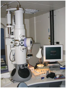
Electron Microscope
The instrument that is used for highly magnified and resolved images of biological or non-biological specimens and objects with a highly energetic beam of electrons is called an electron microscope.
- 1) History of Electron microscope
- 2) Principle
- 3) Construction
- 4) Working of Electron Microscope
- 5) Types of Electron Microscope
- 6) Scanning Electron microscope
- 7) Transmission electron microscope
- 8) Applications and Advantages
- 9) MCQs about Electron Microscope
- 10) Summary:
- 11) You may also like to learn:
History of Electron microscope
In 1931, physicist Ernst Ruska and electrical engineer Max Knoll first invented the electron microscope. This was the practical application of electron microscopy.
Principle
The electron microscope works on the wave behavior of electrons. The beam of electrons is used to magnify and resolve the image by its characteristic wave nature and provide information on the composition, morphology, structure, and other details of the specimen.
Construction
Electron microscope consists of an electron gun, magnetic condenser, magnetic objective, intermediate image projector, and a fluorescent screen. All parts are described below in detail.
Electron gun
The electron gun in the microscope generates the beam of electrons with uniform velocity. Generally, a potential difference of 30 kV to several megavolts is used to highly accelerate the electrons.
Commercially used high voltage microscopes can operate on potential differences up to 1500 kV.
Magnetic condenser
There are two sets of magnetic condensers which focus the electron beam on the specimen. In this, electric and magnetic fields are used for focusing beams of electrons.
Magnetic objective
It is like the objective lens of an optical microscope. Electrons fall on the specimen scattered out from the thicker part and enter the magnetic objective so the first image of the specimen is produced.
Intermediate image projector
This is like the projector lens of an optical microscope. It is a magnetic coil that forms a real intermediate image and the projector lens shows this on a fluorescent screen.
Fluorescent screen
The highly magnified and resolved image of the object or specimen under study is shown on a fluorescent screen.
The special film on which the final image of the specimen is displayed is called an electron micrograph.
Working of Electron Microscope
Stream of electrons are produced and sped up by the electron gun. The electron beam is made to travel through the center of the magnetic condensing lens. These electrons are made as parallel beam and are focused on the objects.
The electrons are sent more in the less thick region of the object and are transmitted less (i.e.,) absorbed up by the denser region of the object.
Therefore, the transmitted electron beam falling over the magnetic objective lens forms a magnified real image. Even more, the image can be amplified by the magnetic projector lens and the final image is obtained on the fluorescent screen.
In order to make a permanent record of the image of the things, the final image can also be obtained on a photographic plate.
Types of Electron Microscope
Today there are two significant kinds of electron microscopic lens used in clinical and biomedical research: the transmission electron microscope (TEM) and the scanning electron microscope (SEM); in some cases, the TEM and SEM are combined in one instrument, the scanning transmission electron microscope (STEM):
TEM: amplifies 50 to ~ 50 million times; the specimen appears flat.
SEM: amplifies 5 to ~ 500,000 times; sharp images of surface area features.
STEM: amplifies 5 to ~ 50 million times; the specimen appears flat.
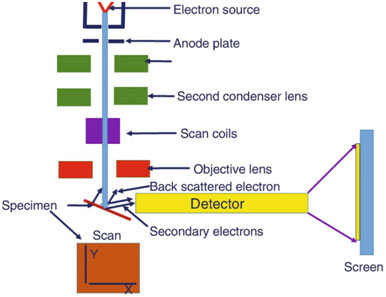
Scanning Electron microscope
Scanning electron microscope is an enhanced design of an electron microscopic lens. SEM is utilized to study the 3-dimensional image of the specimen.
When the accelerated primary electrons strike the specimen, it produces secondary electrons. These secondary electrons are collected by a positiv charged electron detector which in turn provides a 3- dimensional image of the specimen.
Transmission electron microscope
In the TEM, the electrons from the electron gun travel through a condenser lens prior to encountering the specimen, close to the objective lens. The majority of the magnification is accomplished by the objective lens system.
The image is viewed at the base of the column and photographed utilizing film, or more recently a CCD camera, by raising the hinged fluorescent viewing screen.
Applications and Advantages
- Electron microscopes are utilized to investigate the ultrastructure of a wide variety of biological and inorganic specimens consisting of microbes, cells, macromolecules, biopsy samples, metals, and crystals.
- Industrially, electron microscopes are frequently used for quality control and failure analysis.
- The science of microbiology owes its advancement to the electron microscope. Study of microbes like germs, virus and other pathogens have made the treatment of illness really reliable.
Advantages
- It can produce magnification as high as 1, 00,000 times as that of the size of the object.
- The focal length of the microscopic system can differ.
MCQs about Electron Microscope
- 1. What is the purpose of an electron microscope?
- A) To generate electricity
- B) To magnify and resolve images of specimens
- C) To detect magnetic fields
- D) To measure temperature
Answer: B) To magnify and resolve images of specimens
- 2. Who were the inventors of the electron microscope?
- A) Thomas Edison and Nikola Tesla
- B) Albert Einstein and Marie Curie
- C) Ernst Ruska and Max Knoll
- D) Isaac Newton and Galileo Galilei
Answer: C) Ernst Ruska and Max Knoll
- 3. What principle does the electron microscope work on?
- A) Mechanical vibrations
- B) Wave behavior of electrons
- C) Chemical reactions
- D) Gravitational forces
Answer: B) Wave behavior of electrons
- 4. What is the function of the electron gun in an electron microscope?
- A) To generate a beam of electrons
- B) To focus light onto the specimen
- C) To measure temperature
- D) To create a magnetic field
Answer: A) To generate a beam of electrons
- 5. What is the purpose of the magnetic condenser in an electron microscope?
- A) To focus the electron beam on the specimen
- B) To cool down the system
- C) To generate electricity
- D) To produce X-rays
Answer: A) To focus the electron beam on the specimen
- 6. Which type of microscope is used to study the 3-dimensional image of a specimen?
- A) Optical microscope
- B) Electron microscope
- C) Scanning electron microscope (SEM)
- D) Transmission electron microscope (TEM)
Answer: C) Scanning electron microscope (SEM)
- 7. What is the main function of the transmission electron microscope (TEM)?
- A) To amplify surface area features
- B) To produce 3-dimensional images
- C) To amplify up to ~50 million times
- D) To view the base of the column
Answer: D) To view the base of the column
- 8. What are some applications of electron microscopes?
- A) Only used for industrial purposes
- B) Only used for studying metals
- C) Investigating ultrastructure of biological and inorganic specimens
- D) Only used in microbiology
Answer: C) Investigating ultrastructure of biological and inorganic specimens
- 9. What advantage does an electron microscope offer in terms of magnification?
- A) Up to 10x magnification
- B) Up to 100x magnification
- C) Up to 1,000x magnification
- D) Up to 100,000x magnification
Answer: D) Up to 100,000x magnification
- 10. What can vary in the focal length of an electron microscope?
- A) Magnification level
- B) Electron gun intensity
- C) Sample size
- D) Object shape
Answer: A) Magnification level
- 11. Which component of the electron microscope is responsible for producing a magnified real image of the specimen?
- A) Electron gun
- B) Magnetic condenser
- C) Magnetic objective
- D) Fluorescent screen
Answer: C) Magnetic objective
- 12. How do electrons behave in the electron microscope’s magnetic condenser?
- A) They are absorbed by the specimen
- B) They are deflected by electric fields
- C) They are focused on the specimen
- D) They generate X-rays
Answer: C) They are focused on the specimen
- 13. What type of image does the scanning electron microscope (SEM) produce?
- A) Two-dimensional
- B) Three-dimensional
- C) Flat
- D) Magnified
Answer: B) Three-dimensional
- 14. In the transmission electron microscope (TEM), where is the image viewed?
- A) On the electron gun
- B) At the base of the column
- C) On the fluorescent screen
- D) Through the magnetic condenser
Answer: B) At the base of the column
- 15. What is the significance of the scanning transmission electron microscope (STEM)?
- A) It produces flat images
- B) It magnifies up to 50 million times
- C) It provides a three-dimensional view
- D) It combines features of both TEM and SEM
Answer: D) It combines features of both TEM and SEM
- 16. What is one industrial application of electron microscopes?
- A) Studying microorganisms
- B) Quality control and failure analysis
- C) Investigating atomic structure
- D) Producing X-rays
Answer: B) Quality control and failure analysis
- 17. How has the science of microbiology benefited from electron microscopes?
- A) By studying chemical reactions
- B) By examining the behavior of electrons
- C) By advancing treatment of diseases
- D) By measuring gravitational forces
Answer: C) By advancing treatment of diseases
- 18. What does the electron microscope’s fluorescent screen display?
- A) The initial specimen
- B) A two-dimensional image
- C) The intermediate image
- D) The final magnified image
Answer: D) The final magnified image
- 19. What does the electron microscope’s electron micrograph serve as?
- A) A specimen holder
- B) A magnetic condenser
- C) A fluorescent screen
- D) A permanent record of the image
Answer: D) A permanent record of the image
- 20. What range of potential difference is typically used in the electron gun of a microscope?
- A) 1 kV to 10 kV
- B) 10 kV to 100 kV
- C) 100 kV to 1,000 kV
- D) 1,000 kV to 10,000 kV
Answer: B) 10 kV to 100 kV
- 21. What part of the electron microscope is responsible for amplifying the primary electron beam?
- A) Magnetic condenser
- B) Electron gun
- C) Magnetic objective
- D) Intermediate image projector
Answer: D) Intermediate image projector
Summary:
The electron microscope, invented in 1931 by physicist Ernst Ruska and electrical engineer Max Knoll, revolutionized microscopy by allowing highly magnified and resolved images of biological and non-biological specimens. It operates on the wave behavior of electrons, magnifying and resolving images through the characteristic wave nature of electrons.
The construction of an electron microscope involves various components including the electron gun, magnetic condenser, magnetic objective, intermediate image projector, and fluorescent screen. The electron gun generates a beam of electrons accelerated by a potential difference, which is then focused on the specimen by magnetic condensers and objective. The resulting image is projected onto a fluorescent screen for visualization.
There are two main types of electron microscopes: transmission electron microscopes (TEM) and scanning electron microscopes (SEM), each with its unique applications. TEM provides high magnification and resolution, especially for flat specimens, while SEM is ideal for studying three-dimensional images.
These microscopes have advanced fields such as microbiology and industrial quality control. They offer advantages such as high magnification capabilities and adjustable focal lengths, making them indispensable tools in scientific research and industrial applications.